
Diagnostic and therapeutic experience in patients diagnosed with renal neoplasm of low malignant potential after Bosniak Ⅰ-Ⅱ renal cyst decortication(Report of 8 cases)
-
摘要: 目的 探讨术前表现为Bosniak Ⅰ~Ⅱ级肾囊肿,接受肾囊肿去顶降压术后病理确诊为低度恶性潜能多房囊性肾肿瘤患者的影像和病理特征及诊治经验。方法 回顾性分析2018—2023年上海交通大学医学院附属新华医院收治的8例Bosniak Ⅰ~Ⅱ级肾囊肿去顶术后病理确诊低度恶性潜能多房囊性肾肿瘤患者的临床资料,其中男2例,女6例,平均年龄41岁。研究内容包含影像学特征、诊治经过和患者随访情况等。结果 患者术前影像学提示Bosniak Ⅰ~Ⅱ级肾囊肿,术后病理诊断低度恶性潜能多房囊性肾肿瘤。回溯影像典型特征包括囊壁及间隔增厚、强化的高密度灶、内壁毛糙及厚壁样结节等。后续治疗方案包括二期肾部分切除术2例,根治性肾切除术4例,2次治疗窗口期平均2个月(2周~6个月)。患者术后恢复良好,未见复发转移。另2例行积极监测,密切随访未见复发。结论 重视肾脏囊性疾病的术前诊断,提高影像学检查和术中冷冻病理的敏感性,对确诊为低度恶性潜能多房囊性肾肿瘤的患者采取积极治疗,对提升肾脏恶性囊性病变的诊疗具有重要意义。
-
关键词:
- 肾囊肿 /
- 低度恶性潜能多房囊性肾肿瘤 /
- Bosniak分级 /
- 囊液分析
Abstract: Objective To investigate the imaging and pathological characteristics, as well as diagnostic and treatment experience of patients who pathologically confirmed as multilocular cystic renal neoplasm of low malignant potential after Bosniak Ⅰ-Ⅱ renal cyst decortication.Methods A retrospective analysis was conducted on clinical data of 8 patients diagnosed with multilocular renal cyst neoplasm of low malignant potential after Bosniak Ⅰ-Ⅱ renal cystic decortication at Xinhua Hospital Affiliated to Medical College of Shanghai Jiao Tong University between 2018 and 2023. There were 2 males and 6 females with an average age of 41 years old. The analysis included imaging features, diagnostic and treatment procedures, and patient follow-up outcomes.Results The cases were considered as Bosniak Ⅰ-Ⅱ renal cysts, while pathological results confirmed to be multilocular cystic renal tumor of low malignant potential. A retrospective analysis of imaging revealed typical features which included thickening of cyst walls and septa, enhanced high-density masses, and thick-wall nodules. Follow-up treatments included partial nephrectomy in 2 cases, radical nephrectomy in 4 cases, with an average interval of 2 months (2 weeks to 6 months) between the two procedures. Patients exhibited favorable postoperative recovery and no disease progression was observed. Two patients under active monitoring showed no tumor recurrence.Conclusion The key to improve the diagnosis and curative rate of cystic renal masses is paying attention to preoperative imaging study, intraoperative frozen section examination and active follow-up treatment. -

-
表 1 Bosniak Ⅰ~Ⅱ级肾囊肿术后确诊低度恶性潜能多房囊性肾肿瘤患者一般资料
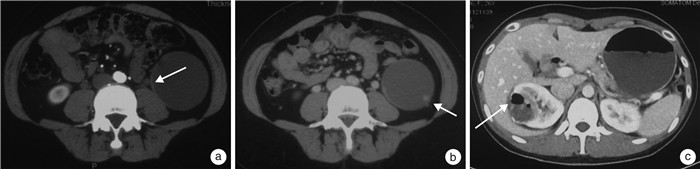
例序 性别 年龄/岁 影像诊断 石蜡病理诊断 回溯影像学特征 1 女 35 右肾Bosniak Ⅰ级肾囊性病变 低度恶性潜能多房囊性肾肿瘤 囊壁多发附壁结节,增强后结节可见早期明显强化,静脉及延迟期强化减低 2 女 41 右肾Bosniak Ⅰ级肾囊性病变 低度恶性潜能多房囊性肾肿瘤 ①右肾内壁厚囊样结节;②右肾下部实质内见边缘稍毛糙、壁稍厚的稍低密度灶 3 男 42 左肾Bosniak Ⅰ级肾囊性病变 倾向低度恶性潜能多房囊性肾肿瘤 ①左肾囊性低密度病变,壁局部不规则增厚伴线样分割影;②囊壁及分隔轻度强化 4 女 53 左肾Bosniak Ⅱ级肾囊性病变 倾向低度恶性潜能多房囊性肾肿瘤 ①壁稍厚的稍低密度灶;②囊内多发间隔,分隔呈交叉分布 5 女 46 右肾Bosniak Ⅰ级肾囊性病变 低度恶性潜能多房囊性肾肿瘤 囊壁及分隔上可见实质性强化软组织结节 6 男 45 右肾Bosniak Ⅱ级肾囊性病变 低度恶性潜能多房囊性肾肿瘤 ①增强可见稍高密度影;②囊壁及分隔上可见实质性强化软组织结节 7 女 32 左肾Bosniak Ⅰ级肾囊性病变 低度恶性潜能多房囊性肾肿瘤 左肾实质内见边缘稍毛糙的稍低密度灶 8 女 35 左肾Bosniak Ⅰ级肾囊性病变 低度恶性潜能多房囊性肾肿瘤 ①囊壁环形钙化伴囊腔内积气;②部分囊壁与相邻附壁粘连伴周围斑片索状影  下载: 导出CSV
-
[1] Chang CC, Kuo JY, Chan WL, et al. Prevalence and clinical characteristics of simple renal cyst[J]. J Chin Med Assoc, 2007, 70(11): 486-491. doi: 10.1016/S1726-4901(08)70046-7
[2] Bosniak MA. The current radiological approach to renal cysts[J]. Radiology, 1986, 158(1): 1-10. doi: 10.1148/radiology.158.1.3510019
[3] Silverman SG, Pedrosa I, Ellis JH, et al. Bosniak classification of cystic renal masses, version 2019: an update proposal and needs assessment[J]. Radiology, 2019, 292(2): 475-488. doi: 10.1148/radiol.2019182646
[4] Boo HJ, Lee JE, Chung SM, et al. The presence of simple renal cysts is associated with an increased risk of albuminuria in young adults[J]. Korean J Intern Med, 2022, 37(2): 425-433. doi: 10.3904/kjim.2020.576
[5] Simms RJ, Ong AC. How simple are 'simple renal cysts'?[J]. Nephrol Dial Transplant, 2014, 29 Suppl 4(Suppl 4): ⅳ106-112.
[6] Bosniak MA. The Bosniak renal cyst classification: 25 years later[J]. Radiology, 2012, 262(3): 781-785. doi: 10.1148/radiol.11111595
[7] Tse JR, Shen LY, Shen J, et al. Prevalence of malignancy and histopathological association of bosniak classification, version 2019 class Ⅲ and Ⅳ cystic renal masses[J]. J Urol, 2021, 205(4): 1031-1038. doi: 10.1097/JU.0000000000001438
[8] Nassir A, Jollimore J, Gupta R, et al. Multilocular cystic renal cell carcinoma: a series of 12 cases and review of the literature[J]. Urology, 2002, 60(3): 421-427. doi: 10.1016/S0090-4295(02)01742-9
[9] Winters BR, Gore JL, Holt SK, et al. Cystic renal cell carcinoma carries an excellent prognosis regardless of tumor size[J]. Urol Oncol, 2015, 33(12): 505.e9-13. doi: 10.1016/j.urolonc.2015.07.017
[10] Moch H, Cubilla AL, Humphrey PA, et al. The 2016 WHO classification of tumours of the urinary system and male genital organs-part A: renal, penile, and testicular tumours[J]. Eur Urol, 2016, 70(1): 93-105. doi: 10.1016/j.eururo.2016.02.029
[11] Oh TH, Seo IY. The role of Bosniak classification in malignant tumor diagnosis: a single institution experience[J]. Investig Clin Urol, 2016, 57(2): 100-105. doi: 10.4111/icu.2016.57.2.100
[12] Qin X, Ye L, Zhang H, et al. Complicated variation of simple renal cyst usually means malignancy: results from a cohort study[J]. World J Surg Oncol, 2014, 12: 316. doi: 10.1186/1477-7819-12-316
[13] Song MG, Lee CH, Kim KA, et al. Simple renal cyst (Bosniak classification type 1 cyst): Is follow-up warranted?[J]. Eur J Radiol Extra, 2009, 72(3): e133-e135. doi: 10.1016/j.ejrex.2009.06.006
[14] Cantisani V, Bertolotto M, Clevert DA, et al. EFSUMB 2020 proposal for a contrast-enhanced ultrasound-adapted bosniak cyst categorization-position statement[J]. Ultraschall Med, 2021, 42(2): 154-166. doi: 10.1055/a-1300-1727
[15] Mensel B, Kühn JP, Kracht F, et al. Prevalence of renal cysts and association with risk factors in a general population: an MRI-based study[J]. Abdom Radiol(NY), 2018, 43(11): 3068-3074. doi: 10.1007/s00261-018-1565-5
[16] Kim NY, Lubner MG, Nystrom JT, et al. Utility of CT texture analysis in differentiating low-attenuation renal cell carcinoma from cysts: a Bi-institutional retrospective study[J]. AJR Am J Roentgenol, 2019, 213(6): 1259-1266. doi: 10.2214/AJR.19.21182
[17] 张宇晗. CT影像组学与机器学习在诊断Bosniak Ⅲ级肾脏囊性病变中的应用[D]. 长春: 吉林大学, 2021.
[18] Yu YL, Ma L, Wang ZH, et al. Renal cell carcinoma presenting as a simple renal cyst: a case report[J]. Mol Clin Oncol, 2017, 6(4): 550-552. doi: 10.3892/mco.2017.1173
[19] Lin CJ, Chen YC, Chen HH, et al. Renal cell carcinoma presenting as a huge simple renal cyst[J]. Med Oncol, 2008, 25(1): 104-106. doi: 10.1007/s12032-007-0049-1
[20] Bas O, Nalbant I, Can Sener N, Firat H, Yeşil S, Zengin K, Yalcınkaya F, Imamoglu A. Management of renal cysts[J]. JSLS, 2015, 19(1): e2014.00097. doi: 10.4293/JSLS.2014.00097
[21] Sadeghi A, Shahrbaf MA, Asadzadeh Aghdaei H, et al. A rare presentation of simple renal cyst: gastrointestinal obstruction[J]. Gastroenterol Hepatol Bed Bench, 2018, 11(4): 359-362.
[22] Gupta M, Sherman A, Rosen DC, et al. Infected renal cyst: elusive diagnosis and percutaneous management[J]. J Endourol Case Rep, 2020, 6(2): 89-91. doi: 10.1089/cren.2019.0128
[23] 潘秀武, 叶剑青, 曲发军, 等. 囊性肾癌的微创保肾手术难点及对策[J]. 临床泌尿外科杂志, 2022, 37(9): 688-692. https://lcmw.whuhzzs.com/article/doi/10.13201/j.issn.1001-1420.2022.09.009
-
图(1)
表(1)
计量
- 文章访问数: 1331
- PDF下载数: 689
- 施引文献: 0