
-
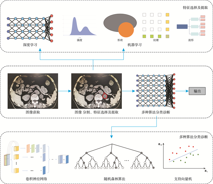
摘要: 近年来,人工智能(artificial Intelligence,AI)凭借其大批量、高维度处理信息的能力,在各个领域展现出快速发展的趋势。在医学影像处理领域,针对肾上腺疾病影像学诊断方面,AI也具备潜在的优势和应用前景。本文将对AI在肾上腺疾病影像学诊断中的研究现状进行综述。Abstract: In recent years, artificial intelligence(AI) has shown a rapid development trend in various fields due to its ability to process large quantities and high-dimensional information. In the field of medical imaging processing, AI has shown great advantages and application prospects in the diagnosis of adrenal diseases. This article will provide a review of the current research status of AI in imaging diagnosis of adrenal diseases.
-
Key words:
- artificial intelligence /
- imaging /
- adrenal diseases /
- diagnosis
-

-
表 1 鉴别肾上腺肿物良恶性
作者 年份 研究目的 方式或方法 模型 研究设计 训练样本量/测试样本量 模型准确率 与放射科医生对比(医生准确率) 参考标准 Elmohr等[2] 2019 鉴别肾上腺肿物的良恶性 CT 单变量逻辑回归随机森林算法 回顾性研究 -/54 76.0%~ 82.0% 有(68.5%) 病理学 Moawad等[3] 2021 鉴别肾上腺肿物的良恶性 CT 二元分类模型 回顾性研究 -/40 71.4% 无 病理学 Shi等[4] 2019 鉴别肾上腺转移瘤和良性肿瘤 CT 支持向量机 回顾性研究 -/265 77.0% 无 组织学 Stanzione等[5] 2021 鉴别肾上腺肿物的良恶性 MRI Extra Trees模型,支持向量机 回顾性研究 -/55 91.0%~ 94.0% 无 病理学+ 随访 Ho等[6] 2019 鉴别肾上腺肿物的良恶性 CT+MRI 因素logistic回归模型 回顾性研究 -/23 80.0% 无 病理学+ 随访 Bi等[7] 2022 鉴别肾上腺肿物的良恶性 CT 深度多尺度相似网络 回顾性研究 229/229 (五折交叉验证) 85.9%~ 89.5% 无 影像学 Singh等[8] 2023 鉴别肾上腺皮质癌和腺瘤 CT 3D Densenet 121模型 回顾性研究 91/91 (五折交叉验证) 87.2%~ 91.0% 无 组织学+ 影像学 Solak等[9] 2023 鉴别肾上腺肿物的良恶性 MRI Abdomen- Caps模型 回顾性研究 122/122 (十折交叉验证) 98.2% 无 影像学 Barstuan等[10] 2020 鉴别肾上腺肿物的良恶性 MRI 人工神经网络,支持向量机 回顾性研究 122/122 (二、五、十折交叉验证) 93.2%~ 98.4% 无 影像学 Cao等[11] 2023 鉴别肾上腺肿物的良恶性 18F-FDG-PET/ CT 多元逻辑回归 回顾性研究 182/121 85.1%~ 88.5% 无 组织病理学 注:CT:计算机断层扫描;MRI:磁共振成像;18F-FDG-PET:[18F]-脱氧葡糖-正电子发射计算机断层显像/计算机断层扫描。  下载: 导出CSV
表 2 鉴别诊断肾上腺腺瘤与嗜铬细胞瘤
作者 年份 研究目的 方式或方法 模型 研究设计 训练样本量/测试样本量 模型准确率 与放射科医生对比(医生准确率) 参考标准 Liu等[23] 2021 鉴别肾上腺腺瘤和嗜铬细胞瘤 MRI 直方图、支持向量机 回顾性研究 40/20 85.0% 无 病理学+ 影像学 Yi等[25] 2018 鉴别肾上腺腺瘤和sPCC CT logistic回归分析 回顾性研究 -/108 85.2%~ 94.4% 无 病理学 Yi等[26] 2018 鉴别肾上腺腺瘤和sPCC CT 多元逻辑回归 回顾性研究 212/53 90.2%~ 96.7% 无 病理学 Altay等[27] 2023 鉴别肾上腺腺瘤、转移瘤和PCC CT logistic回归分析 回顾性研究 -/166 79.5%~ 100.0% 无 病理学+ 内分泌学 Liu等[28] 2022 鉴别肾上腺腺瘤和sPCC CT 多元逻辑回归 回顾性研究 280/280 (五折交叉验证) 83.2%~ 86.4% 无 病理学
下载: 导出CSV
表 3 鉴别肾上腺肿物功能性
作者 年份 研究目的 方式或方法 模型 研究设计 训练样本量/测试样本量 模型准确率 与放射科医生对比(医生准确率) 参考标准 Qi等[36] 2023 鉴别无功能和有功能的腺瘤 CT和增强CT 随机森林、支持向量机、逻辑回归、梯度提升机和极端梯度提升等 回顾性研究 289/54 72.7%~ 83.0% 无 内分泌学 Piskin等[37] 2023 鉴别无功能肿物和CS MRI LASSO回归分析 回顾性研究 69/31 72.1%~ 75.8% 无 内分泌学 Alimu等[38] 2023 鉴别PHA、CS和PCC CT 深度学习模型 回顾性研究 270/105 92.5% 有(80.6%) 病理学+ 内分泌学 Maggio等[43] 2022 鉴别无功能和有功能肾上腺偶发瘤 CT 多元逻辑回归 回顾性研究 -/72 93.8%~ 100.0% 无 内分泌学 Akai等[44] 2020 定位诊断PHA CT logistic回归分析 回顾性研究 -/82 67.1% 无 内分泌学 Sut等[45] 2023 鉴别不同功能的肾上腺偶发瘤 CT K临近算法、支持向量机、神经网络 回顾性研究 96/96 (十折交叉验证) 98.8%~ 99.9% 无 影像学
下载: 导出CSV
表 4 鉴别肾上腺肿物其他方面的应用
第一作者 年份 研究目的 方式或方法 模型 研究设计 训练样本量/测试样本量 模型准确率 与放射科医生对比(医生准确率) 参考标准 Kim等[47] 2023 鉴别肾上腺增生与正常肾上腺 CT 多层感知器,支持向量分类,随机森林分类器,和决策树分类器 回顾性研究 185/123 98.0%~ 99.0% 无 影像学 Saiprasad等[49] 2013 自动分割图像并分类诊断肾上腺疾病 CT 随机森林算法 回顾性研究 10/20 80.0%~ 90.0% 无 影像学 Robinson-Weiss等[50] 2023 自动分割图像并分类诊断肾上腺疾病 CT 深度学习模型 回顾性研究 214/1 051 80.0%~ 91.0% 无 影像学 Kusunoki等[52] 2022 鉴别腺瘤与非腺瘤 CT 深度卷积神经网络 回顾性研究 115/115 (五折交叉验证) 80.0%~ 99.0% 无 病理学+ 随访
下载: 导出CSV
-
[1] 黄健. 中国泌尿外科和男科疾病诊断治疗指南: 2019版[M]. 北京: 科学出版社, 2020: 591-596.
[2] Elmohr MM, Fuentes D, Habra MA, et al. Machine learning-based texture analysis for differentiation of large adrenal cortical tumours on CT[J]. Clin Radiol, 2019, 74(10): 818. e1-818. e7. doi: 10.1016/j.crad.2019.06.021
[3] Moawad AW, Ahmed A, Fuentes DT, et al. Machine learning-based texture analysis for differentiation of radiologically indeterminate small adrenal tumors on adrenal protocol CT scans[J]. Abdom Radiol, 2021, 46(10): 4853-4863. doi: 10.1007/s00261-021-03136-2
[4] Shi B, Zhang GMY, Xu M, et al. Distinguishing metastases from benign adrenal masses: what can CT texture analysis do?[J]. Acta Radiol, 2019, 60(11): 1553-1561. doi: 10.1177/0284185119830292
[5] Stanzione A, Cuocolo R, Verde F, et al. Handcrafted MRI radiomics and machine learning: classification of indeterminate solid adrenal lesions[J]. Magn Reson Imaging, 2021, 79: 52-58. doi: 10.1016/j.mri.2021.03.009
[6] Ho LM, Samei E, Mazurowski MA, et al. Can texture analysis be used to distinguish benign from malignant adrenal nodules on unenhanced CT, contrast-enhanced CT, or in-phase and opposed-phase MRI?[J]. AJR Am J Roentgenol, 2019, 212(3): 554-561. doi: 10.2214/AJR.18.20097
[7] Bi L, Kim J, Su TW, et al. Deep multi-scale resemblance network for the sub-class differentiation of adrenal masses on computed tomography images[J]. Artif Intell Med, 2022, 132: 102374. doi: 10.1016/j.artmed.2022.102374
[8] Singh Y, Kelm ZS, Faghani S, et al. Deep learning approach for differentiating indeterminate adrenal masses using CT imaging[J]. Abdom Radiol(NY), 2023, 48(10): 3189-3194. doi: 10.1007/s00261-023-03988-w
[9] Solak A, Ceylan R, Bozkurt MA, et al. Adrenal lesion classification with abdomen caps and the effect of ROI size[J]. Phys Eng Sci Med, 2023, 46(2): 865-875. doi: 10.1007/s13246-023-01259-y
[10] Barstuǧan M, Ceylan R, Asoglu S, et al. Adrenal tumor characterization on magnetic resonance images[J]. Int J Imaging Syst Tech, 2020, 30(1): 252-265. doi: 10.1002/ima.22358
[11] Cao LX, Zhang DJ, Yang HX, et al. 18F-FDG-PET/CT-based machine learning model evaluates indeterminate adrenal nodules in patients with extra-adrenal malignancies[J]. World J Surg Oncol, 2023, 21(1): 305. doi: 10.1186/s12957-023-03184-6
[12] Fassnacht M, Arlt W, Bancos I, et al. Management of adrenal incidentalomas: European Society of Endocrinology Clinical Practice Guideline in collaboration with the European Network for the Study of Adrenal Tumors[J]. Eur J Endocrinol, 2016, 175(2): G1-G34. doi: 10.1530/EJE-16-0467
[13] 中华医学会内分泌学分会肾上腺学组. 嗜铬细胞瘤和副神经节瘤诊断治疗的专家共识[J]. 中华内分泌代谢杂志, 2016, 32(3): 181-187. doi: 10.3760/cma.j.issn.1000-6699.2016.03.002
[14] Lenders JW, Duh QY, Eisenhofer G, et al. Pheochromocytoma and paraganglioma: an endocrine society clinical practice guideline[J]. J Clin Endocrinol Metab, 2014, 99(6): 1915-1942. doi: 10.1210/jc.2014-1498
[15] Lam AKY. Update on adrenal tumours in 2017 World Health Organization(WHO)of endocrine tumours[J]. Endocr Pathol, 2017, 28(3): 213-227. doi: 10.1007/s12022-017-9484-5
[16] Stuijver DJF, van Zaane B, Feelders RA, et al. Incidence of venous thromboembolism in patients with Cushing's syndrome: a multicenter cohort study[J]. J Clin Endocrinol Metab, 2011, 96(11): 3525-3532. doi: 10.1210/jc.2011-1661
[17] Hönigschnabl S, Gallo S, Niederle B, et al. How accurate is MR imaging in characterisation of adrenal masses: update of a long-term study[J]. Eur J Radiol, 2002, 41(2): 113-122. doi: 10.1016/S0720-048X(01)00443-0
[18] Goldstein RE, O'Neill JA Jr, Holcomb GW 3rd, et al. Clinical experience over 48 years with pheochromocytoma[J]. Ann Surg, 1999, 229(6): 755-764;discussion 764-766. doi: 10.1097/00000658-199906000-00001
[19] Nakajo M, Nakajo M, Fukukura Y, et al. Diagnostic performances of FDG-PET/CT and diffusion-weighted imaging indices for differentiating benign pheochromocytoma from other benign adrenal tumors[J]. Abdom Imaging, 2015, 40(6): 1655-1665. doi: 10.1007/s00261-014-0291-x
[20] 韩文聪, 胡文晖, 陈仕炜, 等. 嗜铬细胞瘤发现方式的临床特征比较分析[J]. 临床泌尿外科杂志, 2023, 38(11): 810-814. https://lcmw.whuhzzs.com/article/doi/10.13201/j.issn.1001-1420.2023.11.002
[21] Jason DS, Oltmann SC. Evaluation of an adrenal incidentaloma[J]. Surg Clin North Am, 2019, 99(4): 721-729. doi: 10.1016/j.suc.2019.04.009
[22] Al-Waeli DK, Mansour AA, Haddad NS. Reliability of adrenal computed tomography in predicting the functionality of adrenal incidentaloma[J]. Niger Postgrad Med J, 2020, 27(2): 101-107. doi: 10.4103/npmj.npmj_156_19
[23] Liu JH, Xue KK, Li SJ, et al. Combined diagnosis of whole-lesion histogram analysis of T1-and T2-weighted imaging for differentiating adrenal adenoma and pheochromocytoma: a support vector machine-based study[J]. J L'association Can Des Radiol, 2021, 72(3): 452-459. doi: 10.1177/0846537120911736
[24] Yan LF, Liu ZY, Wang GY, et al. Angiomyolipoma with minimal fat: differentiation from clear cell renal cell carcinoma and papillary renal cell carcinoma by texture analysis on CT images[J]. Acad Radiol, 2015, 22(9): 1115-1121. doi: 10.1016/j.acra.2015.04.004
[25] Yi XP, Guan X, Chen C, et al. Adrenal incidentaloma: machine learning-based quantitative texture analysis of unenhanced CT can effectively differentiate sPHEO from lipid-poor adrenal adenoma[J]. J Cancer, 2018, 9(19): 3577-3582. doi: 10.7150/jca.26356
[26] Yi XP, Guan X, Zhang YM, et al. Radiomics improves efficiency for differentiating subclinical pheochromocytoma from lipid-poor adenoma: a predictive, preventive and personalized medical approach in adrenal incidentalomas[J]. EPMA J, 2018, 9(4): 421-429. doi: 10.1007/s13167-018-0149-3
[27] Altay C, Başara Akın I, Özgül AH, et al. Machine learning analysis of adrenal lesions: preliminary study evaluating texture analysis in the differentiation of adrenal lesions[J]. Diagn Interv Radiol, 2023, 29(2): 234-243.
[28] Liu HP, Guan X, Xu BB, et al. Computed tomography-based machine learning differentiates adrenal pheochromocytoma from lipid-poor adenoma[J]. Front Endocrinol, 2022, 13: 833413. doi: 10.3389/fendo.2022.833413
[29] Barzon L, Sonino N, Fallo F, et al. Prevalence and natural history of adrenal incidentalomas[J]. Eur J Endocrinol, 2003, 149(4): 273-285. http://www.onacademic.com/detail/journal_1000037586373810_8f91.html
[30] Jr YWF. Diagnosis and treatment of primary aldosteronism: practical clinical perspectives[J]. J Intern Med, 2019, 285(2): 126-148. doi: 10.1111/joim.12831
[31] Duan K, Gomez Hernandez K, Mete O. Clinicopathological correlates of adrenal Cushing's syndrome[J]. J Clin Pathol, 2015, 68(3): 175-186. doi: 10.1136/jclinpath-2014-202612
[32] Young WF, Calhoun DA, Lenders JWM, et al. Screening for endocrine hypertension: an endocrine society scientific statement[J]. Endocr Rev, 2017, 38(2): 103-122. doi: 10.1210/er.2017-00054
[33] Barwick TD, Malhotra A, Webb JA, et al. Embryology of the adrenal glands and its relevance to diagnostic imaging[J]. Clin Radiol, 2005, 60(9): 953-959. doi: 10.1016/j.crad.2005.04.006
[34] Siegel RL, Miller KD, Jemal A, et al. Cancer statistics, 2015[J]. CA Cancer J Clin, 2015, 65(1): 5-29. doi: 10.3322/caac.21254
[35] Korobkin M, Brodeur FJ, Yutzy GG, et al. Differentiation of adrenal adenomas from nonadenomas using CT attenuation values[J]. AJR Am J Roentgenol, 1996, 166(3): 531-536. doi: 10.2214/ajr.166.3.8623622
[36] Qi SL, Zuo YF, Chang RS, et al. Using CT radiomic features based on machine learning models to subtype adrenal adenoma[J]. BMC Cancer, 2023, 23(1): 111. doi: 10.1186/s12885-023-10562-6
[37] Piskin FC, Akkus G, Yucel SP, et al. A machine learning approach to distinguishing between non-functioning and autonomous cortisol secreting adrenal incidentaloma on magnetic resonance imaging using texture analysis[J]. Ir J Med Sci, 2023, 192(3): 1155-1161. doi: 10.1007/s11845-022-03105-8
[38] Alimu P, Fang C, Han YN, et al. Artificial intelligence with a deep learning network for the quantification and distinction of functional adrenal tumors based on contrast-enhanced CT images[J]. Quant Imaging Med Surg, 2023, 13(4): 2675-2687. doi: 10.21037/qims-22-539
[39] Park SY, Park BK, Park JJ, et al. CT sensitivities for large(≥3 cm)adrenal adenoma and cortical carcinoma[J]. Abdom Imaging, 2015, 40(2): 310-317. doi: 10.1007/s00261-014-0202-1
[40] Ozturk E, Onur Sildiroglu H, Kantarci M, et al. Computed tomography findings in diseases of the adrenal gland[J]. Wien Klin Wochenschr, 2009, 121(11-12): 372-381. doi: 10.1007/s00508-009-1190-y
[41] 陈雅童, 赵佳晖, 王永兴, 等. 平扫CT值在常见肾上腺肿瘤鉴别诊断中的应用价值[J]. 临床泌尿外科杂志, 2012, 27(1): 29-32. https://lcmw.whuhzzs.com/article/id/a80a8db7-f4ca-4c74-9a6f-f9374e71389e
[42] Mansour N, Mittermeier A, Walter R, et al. Integration of clinical parameters and CT-based radiomics improves machine learning assisted subtyping of primary hyperaldosteronism[J]. Front Endocrinol(Lausanne), 2023, 14: 1244342. doi: 10.3389/fendo.2023.1244342
[43] Maggio R, Messina F, D'Arrigo B, et al. Machine learning-based texture analysis in the characterization of cortisol secreting vs. non-secreting adrenocortical incidentalomas in CT scan[J]. Front Endocrinol(Lausanne), 2022, 13: 873189. doi: 10.3389/fendo.2022.873189
[44] Akai H, Yasaka K, Kunimatsu A, et al. Application of CT texture analysis to assess the localization of primary aldosteronism[J]. Sci Rep, 2020, 10(1): 472. doi: 10.1038/s41598-020-57427-7
[45] Sut SK, Koc M, Zorlu G, et al. Automated adrenal gland disease classes using patch-based center symmetric local binary pattern technique with CT images[J]. J Digit Imaging, 2023, 36(3): 879-892. doi: 10.1007/s10278-022-00759-9
[46] Kosilek RP, Schopohl J, Grunke M, et al. Automatic face classification of Cushing's syndrome in women-a novel screening approach[J]. Exp Clin Endocrinol Diabetes, 2013, 121(9): 561-564. doi: 10.1055/s-0033-1349124
[47] Kim TM, Choi SJ, Ko JY, et al. Fully automatic volume measurement of the adrenal gland on CT using deep learning to classify adrenal hyperplasia[J]. Eur Radiol, 2023, 33(6): 4292-4302.
[48] Barstuǧan M, Ceylan R, Asoglu S, et al. Adrenal tumor segmentation method for MR images[J]. Comput MethodsPrograms Biomed, 2018, 164: 87-100.
[49] Saiprasad G, Chang CI, Safdar N, et al. Adrenal gland abnormality detection using random forest classification[J]. J Digit Imaging, 2013, 26(5): 891-897. doi: 10.1007/s10278-012-9554-7
[50] Robinson-Weiss C, Patel J, Bizzo BC, et al. Machine learning for adrenal gland segmentation and classification of normal and adrenal masses at CT[J]. Radiology, 2023, 306(2): e220101. doi: 10.1148/radiol.220101
[51] Daye D, Staziaki PV, Furtado VF, et al. CT texture analysis and machine learning improve post-ablation prognostication in patients with adrenal metastases: a proof of concept[J]. Cardiovasc Intervent Radiol, 2019, 42(12): 1771-1776. doi: 10.1007/s00270-019-02336-0
[52] Kusunoki M, Nakayama T, Nishie A, et al. A deep learning-based approach for the diagnosis of adrenal adenoma: a new trial using CT[J]. Br J Radiol, 2022, 95(1135): 20211066. doi: 10.1259/bjr.20211066
-
计量
- 文章访问数: 77
- 施引文献: 0