
Application of cone beam CT calibration based on calcification point in image guided radiotherapy for prostate cancer
-
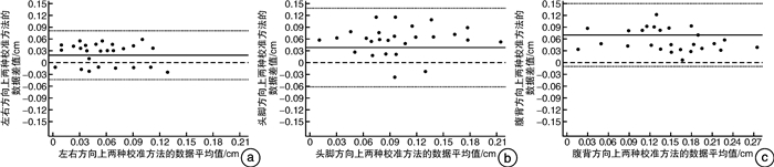
摘要: 目的 以基于骨骼的锥形束CT(CBCT)校准为金标准,评价前列腺癌(PCa)图像引导放疗(IGRT)中基于钙化点的CBCT校准应用的可行性。方法 筛选符合纳排标准的PCa患者30例,接受每周2次治疗前基于骨骼和钙化点的CBCT校准。首先,采用自研算法计算计划CT和首次CBCT3个方向图像上钙化点可探测性分数。图像上钙化点可探测性分数低于0.260分的患者退出试验。之后,以剩余患者基于骨骼的CBCT校准数据为金标准,采用Bland-Altman法分析2种校准方法的数据一致性。结果 4例患者计划CT或首次CBCT图像存在钙化点可探测性分数不达标情况并退出试验。其余26例患者共接受了364次CBCT扫描,其中基于骨骼的校准数据为,左右方向:(0.11±0.07) cm、头脚方向:(0.18±0.13) cm、腹背方向:(0.23±0.22) cm;基于钙化点的校准数据为,左右方向:(0.13±0.11) cm、头脚方向:(0.22±0.19) cm、腹背方向:(0.30±0.25) cm。Bland-Altman分析结果显示,2种校准方法的数据在3个方向上一致性良好。结论 2种校准方法的数据差异较小且一致性良好,在特定条件下基于钙化点的CBCT校准在PCa IGRT中应用具有可行性。Abstract: Objective To evaluate the feasibility of cone beam CT (CBCT) calibration based on calcification point in image guided radiotherapy (IGRT) for prostate cancer (PCa), taking the CBCT calibration based on bone as the gold standard.Methods Thirty patients with PCa who met the inclusion and exclusion criteria were selected and received CBCT calibration based on bone and calcification point before treatment twice a week. Firstly, the self-developed algorithm was used to calculate the detectability scores of calcification points in three directions on the planning CT and the first CBCT images. Patients with calcification point detectability scores less than 0.260 on the images withdrew from the trial. Then, taking the CBCT calibration data based on bone from the remaining patients as the gold standard, the Bland Altman method was used to analyze the data consistency of the two calibration methods.Results The detectability scores of calcification points on planning CT or first CBCT images from 4 patients were not up to standard and they withdrew from the trial. The remaining 26 patients received 364 CBCT scans, of which the calibration data based on bone were as follows, left-right direction: (0.11±0.07) cm, superior-inferior direction: (0.18±0.13) cm, anterior-posterior direction: (0.23±0.22) cm. The calibration data based on calcification point were as follows, left-right direction: (0.13±0.11) cm, superior-inferior direction: (0.22±0.19) cm, anterior-posterior direction: (0.30±0.25) cm. The Bland Altman analysis results showed that the data of the two calibration methods were consistent in three directions.Conclusion The data of the two calibration methods had little difference and good consistency. CBCT calibration based on calcification point was feasible in PCa IGRT under specific conditions.
-
Key words:
- prostate cancer /
- image guided radiotherapy /
- cone beam CT /
- calibration /
- calcification point
-

-
[1] 田龙, 闫洁诚, 胡逸民. 前列腺癌靶区位移影响因素分析[J]. 北京生物医学工程, 2021, 40(4): 406-412. doi: 10.3969/j.issn.1002-3208.2021.04.011
[2] 闫洁诚, 田龙, 胡逸民. 膀胱癌放疗中两种图像引导方法校准精度比较[J]. 北京生物医学工程, 2021, 40(3): 303-308. doi: 10.3969/j.issn.1002-3208.2021.03.013.
[3] Schumacher LD, Dal Pra A, Hoffe SE, et al. Toxicity reduction required for MRI-guided radiotherapy to be cost-effective in the treatment of localized prostate cancer[J]. Br J Radiol, 2020, 93(1114): 20200028. doi: 10.1259/bjr.20200028
[4] Webster A, Appelt AL, Eminowicz G. Image-Guided Radiotherapy for Pelvic Cancers: A Review of Current Evidence and Clinical Utilisation[J]. Clin Oncol(R CollRadiol), 2020, 32(12): 805-816. doi: 10.1016/j.clon.2020.09.010
[5] Murray J, Griffin C, Gulliford S, et al. A randomised assessment of image guided radiotherapy within a phase 3 trial of conventional or hypofractionated high dose intensity modulated radiotherapy for prostate cancer[J]. Radiother Oncol, 2020, 142: 62-71. doi: 10.1016/j.radonc.2019.10.017
[6] 田龙, 席强, 赵鑫, 等. 膀胱癌图像引导放疗中基于骨骼与基于内植标记物锥形束CT校准比较[J]. 中国医学物理学杂志, 2019, 36(6): 647-652. doi: 10.3969/j.issn.1005-202X.2019.06.006
[7] 田龙, 席强, 赵鑫, 等. 膀胱癌图像引导放疗中基于软组织灰度值与基于内植标记物的锥形束CT校准比较[J]. 中国医学物理学杂志, 2018, 35(1): 31-35. doi: 10.3969/j.issn.1005-202X.2018.01.007
[8] Shimada H, Tago M. Prostate cancer dural metastasis resembling a meningioma[J]. Clin Case Rep, 2022, 10(4): e05601.
[9] 田龙, 闫洁诚, 胡逸民, 等. 前列腺癌容积旋转调强放疗中标志物可探测性研究[J]. 国际放射医学核医学杂志, 2021, 45(12): 767-772. doi: 10.3760/cma.j.cn121381-202102024-00123
[10] Preisser F, Heinze A, S Abrams-Pompe R, et al. Impact of positive surgical margin length and Gleason grade at the margin on oncologic outcomes in patients with nonorgan-confined prostate cancer[J]. Prostate, 2022, 82(9): 949-956. doi: 10.1002/pros.24341
[11] Iocolano M, Blacksburg S, Carpenter T, et al. Prostate Fiducial Marker Placement in Patients on Anticoagulation: Feasibility Prior to Prostate SBRT[J]. Front Oncol, 2020, 10: 203. doi: 10.3389/fonc.2020.00203
[12] Ka W, Wong, Ranee, et al. Is there a role for trans-perineal ultrasound imaging of the anal sphincter immediately after primary repair of third degree tears?[J]. Eur J Obstet Gynecol Reprod Biol, 2022, 271: 260-264. doi: 10.1016/j.ejogrb.2022.02.182
[13] Liu ZZ, Tan L, Sharen GW, et al. [Value of Transperineal Ultrasound in Short-term Evaluation of Pelvic Organ Prolapse after Transvaginal Mesh Implantation][J]. Zhongguo Yi Xue Ke Xue Yuan Xue Bao, 2021, 43(6): 892-896.
[14] Kim HY, Choi YH, Lee SJ. Effect of Sedation Anesthesia With Intravenous Propofol on Transrectal Ultrasound-Guided Prostate Biopsy Outcomes[J]. J Korean Med Sci, 2022, 37(15): e115. doi: 10.3346/jkms.2022.37.e115
[15] Takeda S, Fujimoto T, Onda K. Traumatic cervical vertebral artery dissection: A case with cerebral infarct due to newly formed thrombus in the cerebral arteries[J]. Neuropathology, 2020, 40(5): 501-506. doi: 10.1111/neup.12664
[16] O'Neill A, Osman SO, Jain S, et al. Observed high incidence of prostatic calculi with the potential to act as natural fiducials for prostate image guided radiotherapy[J]. Tech Innov Patient Support Radiat Oncol, 2019, 9: 35-40. doi: 10.1016/j.tipsro.2019.01.004
[17] 琚官群, 蔡之平, 张宗勤, 等. "2+2"Trocar分布法在经腹膜外机器人辅助前列腺癌根治术中的应用[J]. 临床泌尿外科杂志, 2021, 36(11): 843-846. http://lcmw.cbpt.cnki.net/WKC/WebPublication/paperDigest.aspx?paperID=765064ee-b036-4e37-b4ee-9b6fa2822194
[18] 邱实, 何龙, 陈鹏, 等. 影响前列腺癌根治性切除术后首次血清PSA水平的因素分析[J]. 临床泌尿外科杂志, 2022, 37(4): 302-305. doi: 10.13201/j.issn.1001-1420.2022.04.012 http://lcmw.cbpt.cnki.net/WKC/WebPublication/paperDigest.aspx?paperID=63bc4f4c-8f4c-47e6-918c-ca7238ab332f
[19] 陈志华, 蒋国松, 阮海龙, 等. 机器人辅助腹腔镜前列腺癌根治术治疗高危局部进展期前列腺癌临床疗效分析[J]. 临床泌尿外科杂志, 2022, 37(2): 123-126. http://lcmw.cbpt.cnki.net/WKC/WebPublication/paperDigest.aspx?paperID=46a706c9-cc53-45d3-9945-a03cf2b9f756
-
 下载:
下载:
图(2)
计量
- 文章访问数: 932
- PDF下载数: 222
- 施引文献: 0