
Comparative analysis of the safety and feasibility of three-dimensional visualization imaging in the surgical treatment of renal tumors in special locations
-
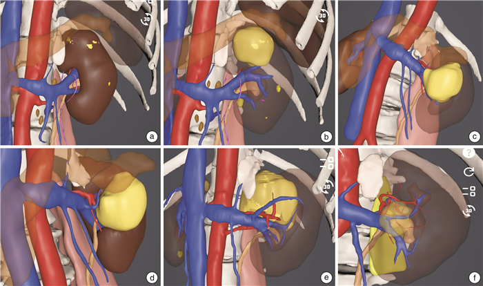
摘要: 目的 比较分析三维可视化成像辅助导引与非三维可视化成像辅助导引手术治疗特殊位置肾肿瘤的安全性和可行性,并分析患者预后情况。方法 回顾性分析2021年3月—2024年5月四川大学华西医院泌尿外科同一医疗组同一术者收治的210例特殊位置肾肿瘤患者的临床资料,根据是否行三维可视化成像导引,分为三维可视化成像辅助导引组(64例)与非三维可视化成像辅助导引组(146例)。比较2组中成功保留肾单位患者比例、是否行血管修补、是否修补集合系统、围术期是否留置输尿管支架、手术时间、肾动脉阻断时间、术中出血量、围术期并发症、术后住院时间及肿瘤有无复发等情况。评价并分析三维可视化成像在特殊位置肾肿瘤手术治疗中的应用价值。结果 210例患者均顺利完成手术,未出现严重术中并发症和围手术期死亡。三维可视化成像辅助导引组有59例(92.2%)患者成功行保留肾单位手术治疗,5例(7.8%)转为根治性肾切除;而非三维可视化成像辅助导引组有75例(51.4%)患者成功行保留肾单位手术治疗,71例(48.6%)患者转为根治性肾切除,差异有统计学意义(P < 0.001)。2组在手术时间[三维可视化成像辅助导引组(125.0±35.0) min、非三维可视化成像辅助导引组(98.6±43.9) min]、集合系统是否打开[三维可视化成像辅助导引组成功保留肾单位患者术中打开集合系统有32例(50.0%)、非三维可视化成像辅助导引组成功保留肾单位患者术中打开集合系统有62例(42.5%)]、术中是否修补血管[三维可视化成像辅助导引组有18例(28.1%)、非三维可视化成像辅助导引组有9例(6.2%)]方面比较差异均有统计学意义(P < 0.001)。结论 应用三维可视化成像辅助导引在特殊位置肾肿瘤保留肾单位手术治疗中具有明显优势。本文的研究为临床实践中采用可视化成像导引在特殊位置肾肿瘤保留肾单位手术治疗提供了强有力的证据,证实了其在处理特殊位置肾肿瘤手术中的重要性和有效性。Abstract: Objective To compare the safety and feasibility of three-dimensional(3D) visualization imaging-guided surgery and non-3D visualization imaging-guided surgery in treating renal tumors located in special areas, as well as to assess patient prognosis.Methods We conducted a retrospective analysis of clinical data from 210 patients with renal tumors in special locations admitted to the Department of Urology at West China Hospital of Sichuan University from March 2021 to May 2024. Patients were divided into two groups: those undergoing surgery with 3D visualization imaging guidance(64 cases) and those without(146 cases). We compared the rates of successful nephron-sparing surgery, vascular repair, collecting system repair, the use of perioperative ureteral stents, operation duration, renal artery occlusion time, intraoperative blood loss, perioperative complications, postoperative hospital stay, and tumor recurrence between the two groups.Results All 210 patients were successfully completed their operations without serious intraoperative complications or perioperative mortality.In the 3D visualization imaging-guided group, 59 patients (92.2%) underwent successful nephron-sparing surgery, but 5 patients (7.8%) were converted to radical nephrectomy. In contrast, the non-3D group achieved successful nephron-sparing surgery in 75 patients (51.4%), but 71 patients (48.6%) were converted to radical nephrectomy. It showed a statistically significant difference (P < 0.001). The average operation time was (125.0±35.0) minutes in the 3D group and (98.6±43.9) minutes in the non-3D group. Additionally, collecting system openings occurred in 32 cases(50.0%) in the 3D group compared to 62 cases(42.5%) in the non-3D group. Vascular repair was performed in 18 cases(28.1%) of the 3D group versus 9 cases(6.2%) in the non-3D group, with both differences being statistically significant(P < 0.001).Conclusion The use of 3D visualization imaging-guided surgery demonstrates significant advantages for nephron-sparing procedures in treating renal tumors in special locations. This study provides robust evidence supporting the clinical application of 3D visualization imaging in nephron-sparing surgeries for renal tumors, confirming its importance and efficacy.
-

-
表 1 2组术前资料分析比较
例(%),X±S 项目 三维可视化成像辅助导引组(64例) 非三维可视化成像辅助导引组(146例) P值 性别 0.005 女 34(53.1) 46(31.5) 男 30(46.9) 100(68.5) 年龄/岁 50.8±12.6 57.5±11.9 < 0.001 肿瘤最大径/cm 4.7±1.6 5.2±2.3 0.106 囊实性 0.555 否 49(76.6) 119(81.5) 是 15(23.4) 27(18.5) R.E.N.A.L.评分/分 0.106 10 25(39.1) 69(47.3) 11 34(53.1) 56(38.4) 12 5(7.8) 21(14.4)  下载: 导出CSV
表 2 2组术中情况分析结果比较
例(%),X±S 项目 三维可视化成像辅助导引组(64例) 非三维可视化成像辅助导引组(146例) P值 手术时间/min 125.0±35.0 98.6±43.9 < 0.001 肾动脉阻断时间/min 27.7±10.1 24.5±6.1 0.036 术中出血量/mL 72.2±126.0 56.3±170.0 0.454 肾周脂肪是否有皂化 1.000 否 59(92.2) 134(91.8) 是 5(7.8) 12(8.2) 是否根治 < 0.001 否 59(92.2) 75(51.4) 是 5(7.8) 71(48.6) 集合系统是否打开 < 0.001 否 32(50.0) 13(8.9) 是 32(50.0) 62(42.5) 是否留置输尿管支架 0.028 否 57(89.1) 141(96.6) 是 7(10.9) 5(3.4) 修补肾静脉 < 0.001 否 46(71.9) 137(93.8) 是 18(28.1) 9(6.2) 肿瘤破裂 0.290 否 61(95.3) 141(96.6) 是 3(4.7) 5(3.4) 中转开放 0.157 否 64(100.0) 142(97.3) 是 0(0) 4(2.7)
下载: 导出CSV
表 3 2组术后情况分析结果比较
例(%),X±S 项目 三维可视化成像辅助导引组(64例) 非三维可视化成像辅助导引组(146例) P值 术后住院时间/d 4.1±1.5 3.7±1.4 0.064 肿瘤复发 0.305 否 64(100.0) 146(100.0) 是 0(0) 0(0) 再次入院 0.518 否 64(100) 145(99.3) 是 0(0) 1(0.7)
下载: 导出CSV
-
[1] Ljungberg B, Albiges L, Abu-Ghanem Y, et al. European association of urology guidelines on renal cell carcinoma: the 2022 update[J]. Eur Urol, 2022, 82(4): 399-410. doi: 10.1016/j.eururo.2022.03.006
[2] 方冬, 杨恺惟, 李学松, 等. 腹腔镜治疗大体积肾癌的临床经验及与开放手术的比较[J]. 现代泌尿外科杂志, 2013, 18(6): 553-556.
[3] Ginsburg KB, Johnson K, Moldovan T, et al. A statewide quality improvement collaborative's adherence to the 2017 American urological association guidelines regarding initial evaluation of patients with clinical T1 renal masses[J]. Urology, 2021, 158: 117-124. doi: 10.1016/j.urology.2021.08.036
[4] Pahernik S, Roos F, Röhrig B, et al. Elective nephron sparing surgery for renal cell carcinoma larger than 4 cm[J]. J Urol, 2008, 179(1): 71-74. doi: 10.1016/j.juro.2007.08.165
[5] Crocerossa F, Carbonara U, Cantiello F, et al. Robot-assisted radical nephrectomy: a systematic review and meta-analysis of comparative studies[J]. Eur Urol, 2021, 80(4): 428-439. doi: 10.1016/j.eururo.2020.10.034
[6] 黄健, 张旭. 中国泌尿外科和男科疾病诊断治疗指南: 2022版[M]. 北京: 科学出版社, 2022.
[7] Kutikov A, Uzzo RG. The R.E.N.A.L. nephrometry score: a comprehensive standardized system for quantitating renal tumor size, location and depth[J]. J Urol, 2009, 182(3): 844-853. doi: 10.1016/j.juro.2009.05.035
[8] 郑荣寿, 陈茹, 韩冰峰, 等. 2022年中国恶性肿瘤流行情况分析[J]. 中华肿瘤杂志, 2024, 46(3): 221-231. doi: 10.3760/cma.j.cn112152-20240119-00035
[9] 李小航, 邹南鑫, 彭程, 等. 机器人辅助腹腔镜保留肾单位手术治疗复杂囊实性肾肿瘤经验与预后分析[J]. 临床泌尿外科杂志, 2024, 39(8): 668-673. https://lcmw.whuhzzs.com/article/doi/10.13201/j.issn.1001-1420.2024.08.003
[10] 黄奕昕, 邹湘鹏, 张志凌, 等. 肾部分切除术后术侧近期肾功能保留的相关因素分析[J]. 中华外科杂志, 2023, 61(12): 1099-1103.
[11] Veccia A, Antonelli A, Uzzo RG, et al. Predictive value of nephrometry scores in nephron-sparing surgery: a systematic review and meta-analysis[J]. Eur Urol Focus, 2020, 6(3): 490-504. doi: 10.1016/j.euf.2019.11.004
[12] Banerjee A, Babu R, Jayaraman D, et al. Preoperative three-dimensional modelling and virtual reality planning aids nephron sparing surgery in a child with bilateral Wilms tumour[J]. BMJ Case Rep, 2024, 17(4): e260600. doi: 10.1136/bcr-2024-260600
[13] Åkerlund J, Ljungberg B, Lundstam S, et al. End-stage renal disease after renal cancer surgery: risk factors and overall survival[J]. Scand J Urol, 2024, 59: 109-116. doi: 10.2340/sju.v59.40322
[14] Peycelon M, Hupertan V, Comperat E, et al. Long-term outcomes after nephron sparing surgery for renal cell carcinoma larger than 4 cm[J]. J Urol, 2009, 181(1): 35-41. doi: 10.1016/j.juro.2008.09.025
[15] 周嘉乐, 吴小荣, 陈勇辉, 等. 腹腔镜下肾部分切除术在cT1期完全内生型肾肿瘤治疗中的临床应用与疗效分析[J]. 临床泌尿外科杂志, 2022, 37(9): 665-668. https://lcmw.whuhzzs.com/article/doi/10.13201/j.issn.1001-1420.2022.09.004
[16] Keskin ET, Can O, Özdemir H, et al. Risk factors of open surgery conversion in laparoscopic partial nephrectomy to achieve nephron sparing[J]. Ann Surg Oncol, 2024, 31(6): 3880-3886. doi: 10.1245/s10434-024-15106-1
[17] 马权, 陈永良, 何建松, 等. 三维可视化重建联合肾周冰水降温技术在腹腔镜肾部分切除术中的应用[J]. 临床泌尿外科杂志, 2023, 38(8): 584-588, 595. https://lcmw.whuhzzs.com/article/doi/10.13201/j.issn.1001-1420.2023.08.004
[18] Rosiello G, Larcher A, Fallara G, et al. The impact of intraoperative bleeding on the risk of chronic kidney disease after nephron-sparing surgery[J]. World J Urol, 2021, 39(7): 2553-2558. doi: 10.1007/s00345-020-03504-5
[19] Porpiglia F, Checcucci E, Amparore D, et al. Three-dimensional augmented reality robot-assisted partial nephrectomy in case of complex tumours(PADUA ≥10): a new intraoperative tool overcoming the ultrasound guidance[J]. Eur Urol, 2020, 78(2): 229-238. doi: 10.1016/j.eururo.2019.11.024
-
图(1)
表(3)
计量
- 文章访问数: 571
- PDF下载数: 44
- 施引文献: 0